It has been validated through clinical studies, demonstrating high accuracy in detecting visual field defects comparable to traditional in-clinic methods. There are more than 10 peer-reviewed validation study showing comparable output to gold-standard Humphrey Field Analyzer and confirmed by 6 independent validation studies to-date. The system utilizes advanced algorithms to ensure reliable results, even in non-clinical environments, making it a trusted tool for vision health professionals worldwide.
Yes, the Rapid Online Vision System is a highly effective tool for managing glaucoma. It supports a variety of visual field tests, including threshold testing and screening assessments, to detect early signs of glaucoma and monitor changes in the visual field over time. It incorporates advanced progression analysis tools, enabling eye care professionals to track and analyze changes in a patients visual field with precision. These tools help identify patterns of vision loss, evaluate the effectiveness of treatments, and guide adjustments to management plans for optimal patient outcomes.
The system also offers telehealth functionality, allowing patients to perform visual field tests remotely. This makes it possible for healthcare providers to monitor glaucoma progression and provide timely interventions without requiring frequent in-clinic visits. This feature is especially valuable for patients in remote locations or with limited mobility.
Additionally, Rapid Online Vision System is a registered medical device, meeting international standards for safety and performance. It holds certifications including the CE Mark (Europe), UKCA (United Kingdom), TGA (Australia), MedSafe (New Zealand), and CDSCO (India), ensuring its reliability and compliance with stringent medical regulations.
By combining advanced testing capabilities, progression analysis, telehealth functionality, and international certifications, It provides comprehensive and convenient glaucoma management for both patients and healthcare providers.
Yes, the Rapid Online Vision System is designed to be intuitive and user-friendly for both doctors and patients. Patients can easily access the system via a web browser, follow simple on-screen instructions, and complete visual field tests in just a few minutes, even from the comfort of their homes.
For doctors, the system provides seamless access to test results through detailed, easy-to-interpret reports. These reports are designed to support clinical decision-making, enabling healthcare professionals to track patient progress efficiently and incorporate the system into their existing workflows. This dual usability ensures that it enhances both patient engagement and clinical efficiency.
Yes, it is designed to seamlessly integrate into clinical workflows. Its comprehensive reporting features allow ophthalmologists and optometrists to easily access, review, and share test results with patients or other healthcare providers, improving collaboration and streamlining the management of visual field data.
It uses a Bayesian predictor for returning the visual field threshold given a patient’s response matrix to spots of different brightness. It can achieve results equivalent to standard equipment in 1.5 minutes less test time with a similar level of accuracy. This approach is similar to that adopted by Humphrey in their testing technique called SITA but it has been optimised to return fast and reliable outcomes.
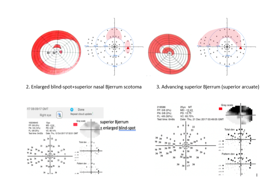
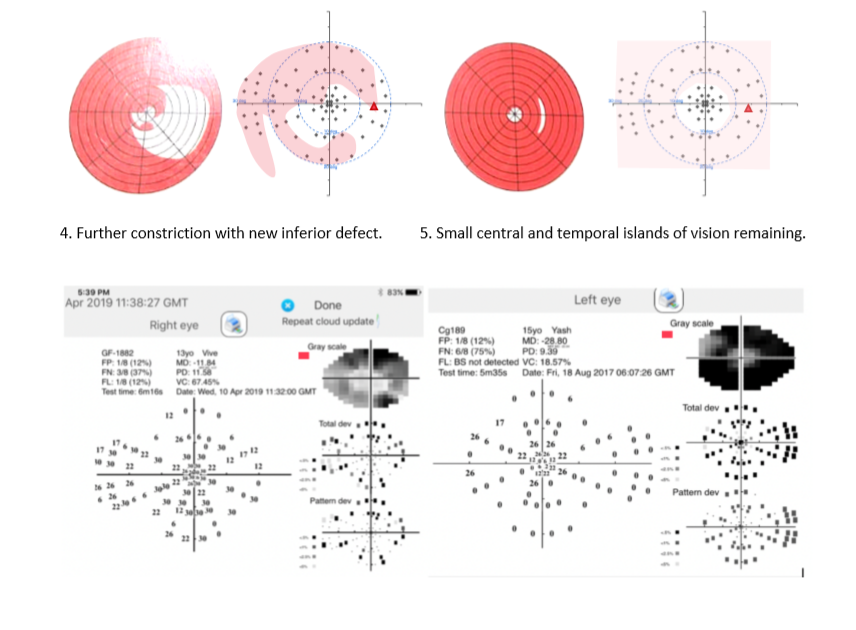
YES, the MRF uses a blind-spot monitor to gauge fixation accuracy. This procedure is common to most perimeters and it starts the test by locating the person’s blind spot. Thereafter it shows spots in the blind spot with the expectation that they should not be seen if fixation is accurate. In our printout the blind spot is shown as a large gap in the graphic. I have shown it as a RED triangle in the below cases to help visualise its location. In some patients the blind spot cannot be detected reliably, and this is recorded on the printout.
Current generation of perimeters do not include enhancements learned over the past 30 years. In fact, the grid and test approaches (spot size) are the same as those of the past. In this aspect nothing has changed, and we are still using the dated approaches of Goldmann.
There is now a large body of evidence that shows that the standard (24-2 and 30-2) grids under-sample the macula, the region of highest density of retinal ganglion cells, so in many cases clinicians need to put patients through a second test, the 10-2. What a pain for both the patient and the clinician.
24-2C has been optimised for glaucoma but will they cope with the more common causes of central vision loss such as AMD or macular oedema?
Since vision loss varies with disease, Rapid Online Vision System is optimised to include four grid patterns specific to glaucoma, maculopathy/AMD, diabetes and neural diseases.
The novel implementations in these designs are a spot size that increases with eccentricity to reduce threshold variability: an expanding test grid to define edges of vision loss and a Bayes threshold predictor with neighbourhood logic to return fast, reliable thresholds (2-3 minutes).
Moreover, Rapid Online Vision System includes three forms of acuity testing that identify refractive/optical changes, retinal or brain losses.
At present the Rapid Online Vision System uses a blind-spot monitor to detect fixation loss (see above point 5).
For those who are interested, an independent clinical trial performed in Sydney by Dr Stuart Grahams group reached the below conclusion
Please refer to this report “Schulz et al. Performance of iPad-based threshold perimetry in glaucoma and controls. Clinical and Experimental Ophthalmology 2018; 46: 346– 355 doi: 10.1111/ceo.13082”
Yes, the Rapid Online Vision System has been used extensively by Dr Viney Gupta and his team (Dept of Ophthalmology (R.P.C) AIIMS, New Delhi). His findings on 21 glaucoma adults have been published as part of a large clinical trial including 39 cases tested in Cambridge UK and his recent work on 43 eyes of 26 children with congenital glaucoma are presently being analysed.
Low Contrast Low Luminance (LLLC) acuity test is great for detecting disorder of retinal pathology. Macular function can be assessed using LLLC in adults with Type 2 Diabetes Mellitus.
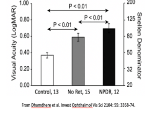
As per research, Smith–Kettlewell Institute Low Luminance (SKILL) Card demonstrates vision function changes in diabetes even in the absence of clinically evident retinopathy. Diabetic retinopathy leads to a further increase in SKILL score, while high contrast VA remains unchanged and standard contrast sensitivity also shows marginal effects.
The research is titled “Assessment of Macular Function Using the SKILL Card in Adults with Type 2 Diabetes Mellitus” and is available on request.
Low Luminance Low Contrast (LLLC – labelled ‘Low contrast Snellen’) is expected to be 6/12 or better for healthy macula and retinal function. The Smith–Kettlewell Institute Low Luminance (SKILL) Card has been used in patients with type 2 diabetes mellitus (T2DM) and been found to have a high sensitivity to abnormal retinal function caused by diabetes. The LLLC charts can be used to evaluate the impact of reduced contrast and reduced luminance on visual acuity.
LLLC vision testing has been found to predict subsequent acuity loss in an aged population as per research. Tests of low contrast spatial vision are strong predictors of early retinal dysfunction. These findings have implications for clinical trials, clinical management, and acceptance of these measures into clinical practice.
The research is titled “Low contrast vision function predicts subsequent acuity loss in an aged population: the SKI study” and the research paper is available on request.
Visual Field Testing
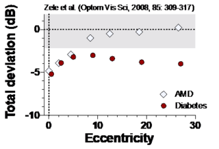
Patients with early diabetes show a loss of
sensitivity across the visual field.
Low Contrast Acuity Tests
The specific tests for Diabetes related eye issues are:
Full Visual Field Test (3.5 minutes)
Macula 10-2 test
Low Luminance Low Contrast (for Diabetes)
High Contrast Acuity